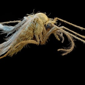
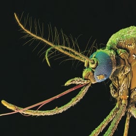
Subcutaneous Dirofilariosis
Diagnosis
Adult worms
As Dirofilaria repens is mainly located in subcutaneous tissues, small, painless nodules with varying location may be investigated surgically. The samples can be preserved in 5% formaldehyde solution or alcohol.
Adult worms are long and whitish, without any grossly obvious identifying features. Males are about 5-7 cm and females are about 10-17 cm in length. Under magnification using a stereomicroscope (x10 to x40), male worms can be observed to possess a series of small papillae arranged in a "v" shape and two unequal spicules at their caudal end.
Microfilariae
For the detection of microfilariae, blood samples should be prevented from coagulation using EDTA or heparin. These samples need to be processed in order to remove other cellular components. This can be performed using the modified Knott’s technique (see also heartworm diagnosis). Microfilariae are 207-360 µm long and 5-8 µm wide. There are overlaps with the sizes of other microfilariae, so confirmation of identity is a specialist skill. Immunological tests have been described that may be used to discriminate between D. immitis and D. repens infections using a blood sample.
Table 1: Details on the differential diagnosis of microfilariae in the blood of dogs (after Deplazes, 2006)
Criteria | D. immitis | D. repens | Acanthocheilonema reconditum | Dipetalonema dracunculoides* | Dipetalonema (Cercopithifilaria) grassi** |
sheath | missing | missing | missing | missing | present, smooth and delicate7 |
approx. length (µm) in stained preparation | 205-2831 | 260-3081 | 213-2402 | 246-2583 | >5507 (660)8 |
mean | <2701 | >2701 | not specified | 2523 | not specified |
width (µm)4 | 5.0-6.5 | 6.0-8.0 | 4.0-5.0 | 5.0-6.0 | 5-15 |
front end | conical | blunt | blunt | conical | blunt |
posterior end | straight | hook-shaped bent | hook-shaped bent (only 30-40%) | straight | |
evidence of acid phosphatase5 | at excretion and anal porus | at anal porus | diffusely distributed | one spot each on the inner body and the anal porus; circle around excretion porus6 | |
molecularbiol. differentiation | PCR | PCR | PCR | PCR |
1Bucklar et al. (1998); 2Whiteley (1988); 3Olmeda-Garcia and Rodriguez-Rodriguez (1994); 4Ducos de Lahitte (1990); 5method: Chalifoux and Hunt (1971); 6Ortega-Mora et al. (1989); 7Euzeby (1981); 8Tarello (2004)
*syn. Acanthocheilonema dracunculoides
**syn. Acanthocheilonema or Cercopithifilaria grassii; more frequently found on dermal tissue
Further information
- Bucklar H, Scheu U, Mossi R, et al.: [Is dirofilariasis in dogs spreading in south Switzerland?] Schweiz Arch Tierheilkd. 1998, 140, 255-60 [in German]
- Chalifoux L, Hunt RD: Histochemical differentiation of Dirofilaria immitis and Dipetalonema reconditum. J Am Vet Med Assoc. 1971, 5, 601-5
- Ducos de Lahitte J: Epidemiologie des filarioses en France. Pratiq Med Chirurg Anim Compag. 1990, 25, 305-10
- Deplazes, P.: [Helminthoses of dogs and cats.] In: Schnieder T (ed.): Veterinärmedizinische Parasitologie. 6th edn., 2006, Parey in MVS, Stuttgart, pp 444-520 [in German]
- Euzeby J: Diagnostic Expérimental des Helminthoses animales. 1981, ed. „Informations Techniques des Services Vétérinaires“, Ministére de l’Agriculture, Paris, France
- Olmeda-Garcia AS, Rodriguez-Rodriguez JA: Stage-specific development of a filarial nematode (Dipetalonema dracunculoides) in vector ticks. J Helminthol. 1994, 68, 231-5
- Ortega-Mora LM, Gomez-Bautista M, Rojo-Vazquez FA: The acid phosphatase activity and morphological characteristics of Dipetalonema dracunculoides (Cobbold, 1870) microfilariae. Vet Parasitol. 1989, 33, 187-90
- Tarello W: Identification and treatment of Dipetalonema grassii microfilariae in a cat from central Italy. Vet Rec. 2004, 155, 565-6
- Whiteley HE: Your diagnostic protocol for Dirofilaria immitis infection in dogs. Vet Med. 1988, 83, 328-45